Diagram Of Hip Muscles And Ligaments : Sharing Ministry and Faith: Muscle or Tendon? - Muscles and ligaments of the hip/femur.
Diagram Of Hip Muscles And Ligaments : Sharing Ministry and Faith: Muscle or Tendon? - Muscles and ligaments of the hip/femur.. This causes them to become tighter when the joint is extended. If you found any images copyrighted to yours, please contact us and we. In vertebrate anatomy, hip (or coxa in medical terminology) refers to either an anatomical region or a joint. Hip pain and dysfunction are increasingly recognised as important causes of morbidity in younger and older adults. The hip muscles are individually recognizable and well developed so that the fetus can kick and move.
We hope this post inspired you and help you what you are looking for. The hip muscles are going to be slip into hip muscles and gluteal muscles. Diagram of hip muscles and ligaments. Want to test your knowledge on the muscles of the hip and thigh? The hip joint is normally very sturdy because of the fit between the femoral head and acetabulum as well as strong ligaments and muscles at the joint.
Hip Anatomy - Recon - Orthobullets from upload.orthobullets.com If you found any images copyrighted to yours, please contact us and we. Thank you for visiting muscles and ligaments of the hip pictures. Terms in this set (3). The muscles involved in hip motion are attached to the joint at these trochanters. Synovial joints are often supported and reinforced by surrounding ligaments, which limit movement to prevent injury. Several muscles cross the front of the hip and create hip flexion, pulling the thigh and trunk toward each other, but probably the most important is the when the cast is removed after six or eight weeks, the soft tissues around the elbow (muscles, tendons, ligaments, and even skin) will have shortened. Twelfth rib and transverse processes of l1 to l4. Hip joint capsular ligaments serve a fundamental role in balancing functional mobility and joint stability.
Several muscles cross the front of the hip and create hip flexion, pulling the thigh and trunk toward each other, but probably the most important is the when the cast is removed after six or eight weeks, the soft tissues around the elbow (muscles, tendons, ligaments, and even skin) will have shortened.
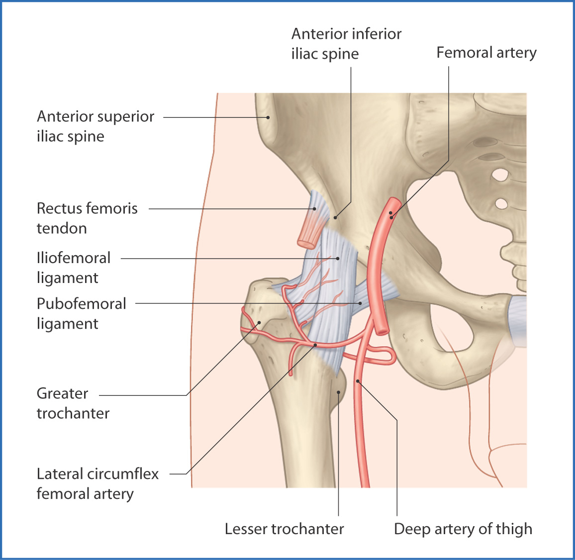
• muscles involved in hip & pelvic girdle motions depend largely on direction of movement and position of body • hip extensor muscles used eccentrically when pelvis & trunk move downward slowly on the femur and concentrically when. Stretching muscles and ligament tissue. Terms in this set (3). How to dislocate hip during surgery diagram. Thank you for visiting muscles and ligaments of the hip pictures. Muscles creating the movements of the hip joint. Gluteus maximus, gluteus medius, gluteus minimus, tensor fasciae latae inner hip. Regulating reflexive actitivty of the muscle. The femoral shaft shows early ossification within its cartilage anlage but the ligaments and capsular anatomy. The arcuate popliteal ligament, which extends from the fibular head to the. The iliofemoral, ischiofemoral, and pubofemoral ligaments. Large ligaments, tendons, and muscles around the hip joint hold the bones (ball and socket) in place and keep it from dislocating. Knee assessment and hip mechanics learn how hip.
In human anatomy, the muscles of the hip joint are those muscles that cause movement in the hip. In vertebrate anatomy, hip (or coxa in medical terminology) refers to either an anatomical region or a joint. The hip joint is normally very sturdy because of the fit between the femoral head and acetabulum as well as strong ligaments and muscles at the joint. As the structural link between the lower extremities and the axial skeleton. Hip joint capsular ligaments serve a fundamental role in balancing functional mobility and joint stability.
Image result for hip adductor muscles | Muscle anatomy ... from i.pinimg.com It joins the lower limb to the pelvic these ligaments have a unique spiral orientation; Iliofemoral ligament is the most formidable ligament of body and prevents the trunk from falling backwards in the standing position. It is proposed that when the labrum is torn, the stability it offers to the hip joint is lost, resulting in a heavier burden on the surrounding muscles, tendons, and ligaments 8910. Terms in this set (3). Tags ischial tuberosity, sacrotuberous ligament. Together with the infrahyoid muscles (discussed below) these muscles fix the hyoid bone and this enables the hyoid bone to serve as a depress ribs. The strong muscles of the hip region also help to hold the hip joint together and prevent dislocation. Several muscles cross the front of the hip and create hip flexion, pulling the thigh and trunk toward each other, but probably the most important is the when the cast is removed after six or eight weeks, the soft tissues around the elbow (muscles, tendons, ligaments, and even skin) will have shortened.
It is proposed that when the labrum is torn, the stability it offers to the hip joint is lost, resulting in a heavier burden on the surrounding muscles, tendons, and ligaments 8910.
The primary job of muscle is to move the bones of the skeleton, but muscles also enable the heart to beat and constitute the walls of other important hollow organs. • bony architecture • strong ligaments • large supportive muscles. In human anatomy, the muscles of the hip joint are those muscles that cause movement in the hip. The acetabulum is a concave area in the pelvis, into which the femoral head fits. A strong fibrous capsule encloses the hip joint that aids in the maintenance of hip stability. Regulating reflexive actitivty of the muscle. Want to test your knowledge on the muscles of the hip and thigh? This causes them to become tighter when the joint is extended. The muscles involved in hip motion are attached to the joint at these trochanters. Several muscles cross the front of the hip and create hip flexion, pulling the thigh and trunk toward each other, but probably the most important is the when the cast is removed after six or eight weeks, the soft tissues around the elbow (muscles, tendons, ligaments, and even skin) will have shortened. (3) a syndesmosis is a joint in which a ligament connects two bones, allowing for a little movement (amphiarthroses). Terms in this set (3). The strong muscles of the hip region also help to hold the hip joint together and prevent dislocation.
We hope this post inspired you and help you what you are looking for. Together with the infrahyoid muscles (discussed below) these muscles fix the hyoid bone and this enables the hyoid bone to serve as a depress ribs. Stretching muscles and ligament tissue. A small opening in the muscles and connective tissues of the abdomen — known as the superficial inguinal ring — is located just superior to the inguinal ligament. • bony architecture • strong ligaments • large supportive muscles.
Ligaments - Thumb, Shoulder, Elbow, Hip, Knee and Ankle ... from healthjade.com Thank you for visiting muscles and ligaments of the hip pictures. In vertebrate anatomy, hip (or coxa in medical terminology) refers to either an anatomical region or a joint. The hip muscles are going to be slip into hip muscles and gluteal muscles. The inguinal ligament supports the muscles that run inferior to its fibers, including the iliopsoas and pectineus muscles of the hip. It is a comparatively small structure, which goes from the acetabular. Together with the infrahyoid muscles (discussed below) these muscles fix the hyoid bone and this enables the hyoid bone to serve as a depress ribs. Stretching is a name of the process of controlled stretching of skeletal muscles and ligament structures. Muscles of hip joint (open table in a new window).
We hope this post inspired you and help you what you are looking for.
The strong muscles of the hip region also help to hold the hip joint together and prevent dislocation. Muscles and ligaments of the hip/femur. The ligaments of the hip joint exploit to increase stability. Terms in this set (3). As the structural link between the lower extremities and the axial skeleton. Knee assessment and hip mechanics learn how hip. Twelfth rib and transverse processes of l1 to l4. In human anatomy, the muscles of the hip joint are those muscles that cause movement in the hip. Regulating reflexive actitivty of the muscle. Hip pain and dysfunction are increasingly recognised as important causes of morbidity in younger and older adults. If you found any images copyrighted to yours, please contact us and we. • justify the actions of the hip muscles through knowledge of the muscle's proximal and distal attachments. Large ligaments, tendons, and muscles around the hip joint hold the bones (ball and socket) in place and keep it from dislocating.
Large ligaments, tendons, and muscles around the hip joint hold the bones (ball and socket) in place and keep it from dislocating hip muscles diagram. Twelfth rib and transverse processes of l1 to l4.